3D Spin Label Imaging

Achieving nanometer-scale imaging of crucial biomolecules like individual virus particles and protein molecules holds immense promise for advancing both structural biology and targeted drug delivery. Conventional methods for structural determination of the relevant biomolecules rely on indirect measurements through the spectroscopic properties of spin labels like DEER. However, limitations arise when multiple spin labels are used, making the inference of the accurate geometry from DEER challenging.
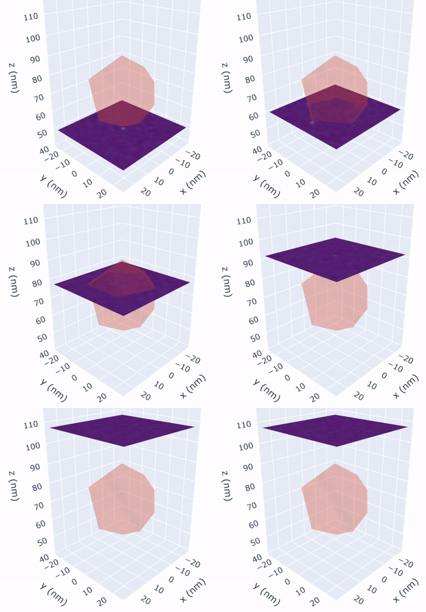
With direct Fourier imaging employing single spin labels at nanometer resolution, a comprehensive image of the molecule can be obtained. Our goal is to devise an imaging protocol that integrates spin labeling techniques to enable this breakthrough.

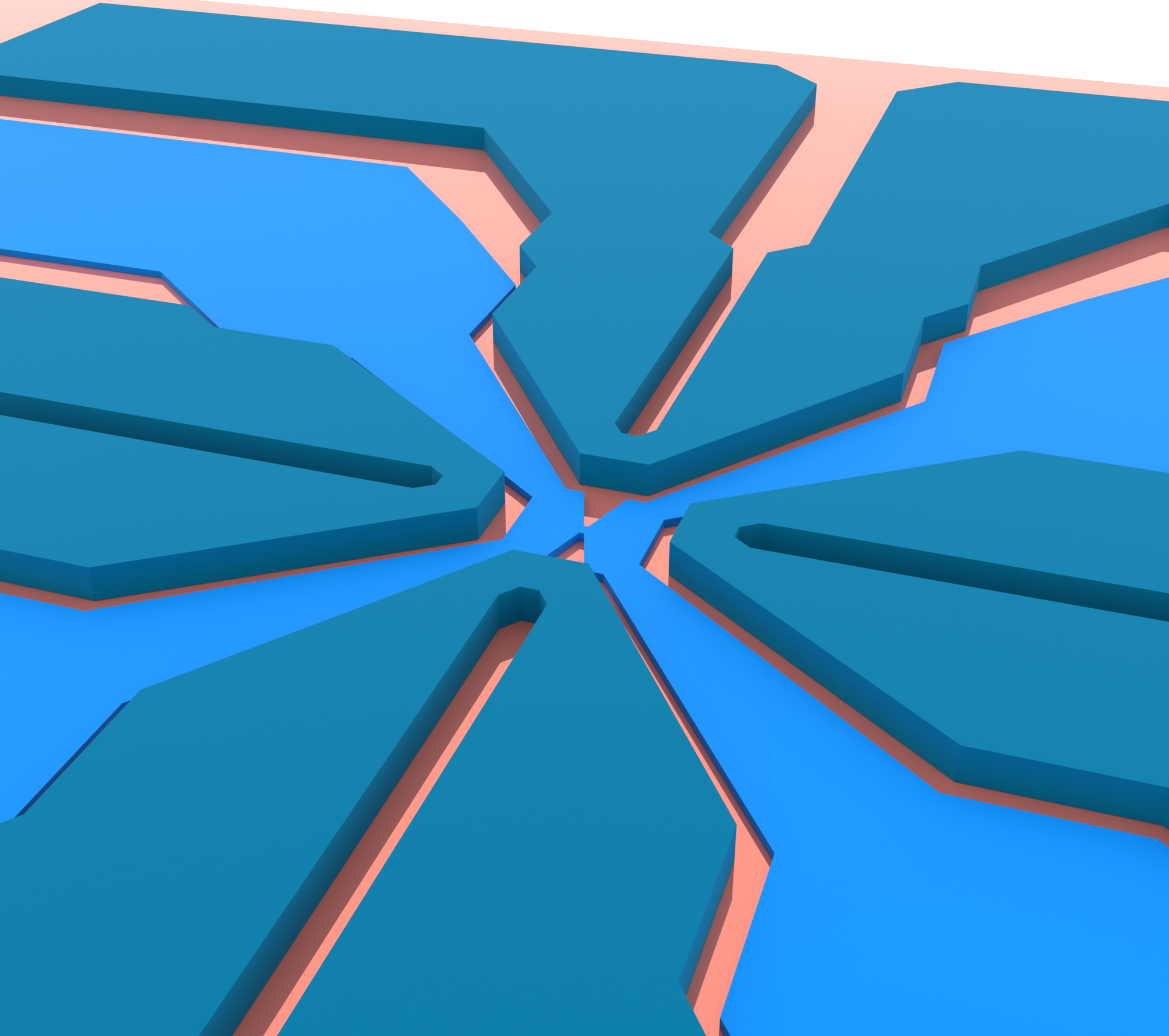
This project involves the design, characterization and fabrication of the next-generation magnetic field gradient sources that operate at the X band (∼10 GHz). These devices would generate strong uniform magnetic field gradients in all 3 spatial directions which are crucial for a 3D Fourier transform MRI with nanometer resolution. These devices, along with ultrasensitive spin detection and high-fidelity quantum control, can be used to image electron spin labels attached to the sample under study.
Another crucial aspect of this project is divising sample preparation protocols to encapsulate the biomolecule or virus particle of interest within a glassy matrix (required as a cryoprotectant), which would in turn be attached to the tip of our ultrasensitive force sensors i.e., the silicon nanowires.
Given the sparse nature of the spin density in this research problem, more sophesticated acquisition methods such as compressive sensing can be used to significantly speed up the imaging.